Features
The JCM-7000 Benchtop Scanning Electron Microscope is designed based on a key concept of "Easy-to-use SEM with seamless navigation and live analysis". The JCM-7000 incorporates three innovative functions; "Zeromag" for smooth transition from optical to SEM imaging, "Live Analysis" for finding constituent elements for an image observation area, and "Live 3D" for displaying a reconstructed live 3D image during SEM observation.
When you place the JCM-7000 next to an optical microscope, further-faster and more-detailed foreign material analysis and quality control can be made.

|
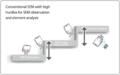
Conventional SEM In conventional SEM operation, SEM imaging and elemental analysis were separated (not seamless). |
|
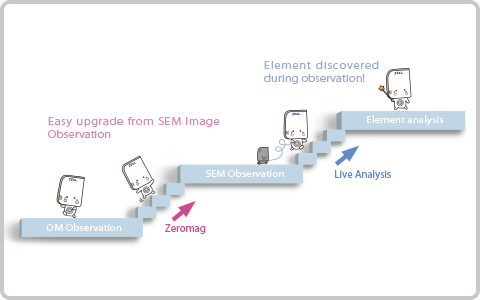
JCM-7000 With "Zeromag", the JCM-7000 enables seamless operation from optical to SEM imaging. Also with "Live Analysis", elemental analysis by EDS can be made during SEM image observation. |
Contaminant analysis
Easy to detect foreign material
Easy to identify elemental composition
【Example】Analysis of black foreign material adhered on surface of food product
|
OM image
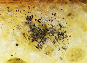 OM observation shows a black powder on specimen surface.
|
|
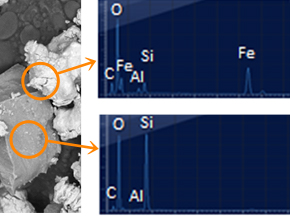
SEM Image (Backscattered electron compositional image 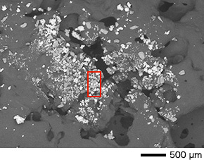 SEM image from the same field of view (FOV) shows particles with different contrast indicating different compositions. SEM image from the same field of view (FOV) shows particles with different contrast indicating different compositions. |
|
SEM Image (Backscattered electron compositional image) Enlarging the area of interest accesses instant live EDS analysis with main elements identified. |
Quality control
Observe detailed surface structures with high resolution and large depth of field not possible with OM imaging.
【Example】Analysis of black foreign material adhered on surface of food product
|
OM image
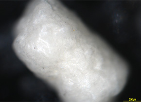 In OM image, it is difficult to see the distribution of the lubricant on the granule surface and quality of its adhesion. |
|
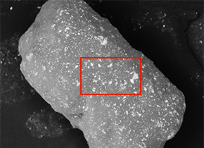
SEM Image (Backscattered electron compositional image)
The superior depth of focus provided withSEM imaging over OM imaging along with the compositional contrast provided withthe backscattered electron detectoclearlyshows the distribution of the lubricant on the surface of the granule. |
|
SEM Image (Backscattered electron compositional image) 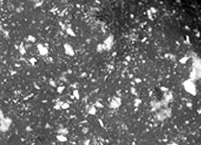 Condition of the lubricant's adhesion can be observed with higher magnification. |
|
Conventional SEM In conventional SEM operation, SEM imaging and elemental analysis were separated (not seamless). |
|
JCM-7000 With "Zeromag", the JCM-7000 enables seamless operation from optical to SEM imaging. Also with "Live Analysis", elemental analysis by EDS can be made during SEM image observation. |